
EVALUATION OF IODINE AND CALCIUM CT NUMBER IN DUAL-ENERGY COMPUTED TOMOGRAPHY (DECT) USING A FABRICATED IN-HOUSE PHANTOM
DOI:
https://doi.org/10.11113/jurnalteknologi.v88.25139Keywords:
CT number, dual energy CT, phantom, quality control, IndoQCTAbstract
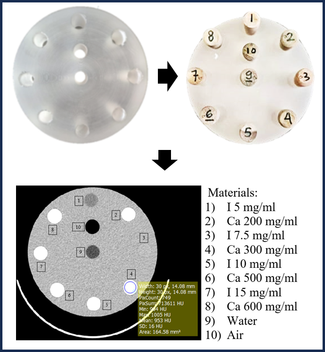
The aim of this study was to develop an in-house phantom with inserts of iodine and calcium materials for evaluating the accuracy of CT numbers from virtual monochromatic image (VMI) of DECT. The phantom was made from polyester resin (PESR) mixed with methyl ethyl ketone peroxide (MEKP) as catalyst in a ratio of 300:1, with a 16 cm diameter and 5 cm height. The phantom had 10 holes, each with a diameter of 1.5 cm. Each hole was filled with water, air, iodine (with various concentrations of 5, 7.5, 10, and 15 mg/ml), and calcium (with various concentrations of 200, 300, 500, and 600 mg/ml). The phantom was scanned by an Ultrafast kV Switching DECT with a tube voltage of 80/140 kV, slice thickness of 5 mm, rotation time of 0.5 s, and tube current variations (200, 250, 300, 335, and 370 mA). Images were reconstructed to a VMI at 50, 70, and 100 keV. The CT number accuracy of VMI images was measured using IndoQCT software. Absolute percentage error (APE) was calculated from measured CT numbers and standard phantom. We found that the changes in CT numbers of iodine and calcium showed the same trend. The CT numbers for both iodine and calcium slightly increased with higher tube currents. The CT number decreased by 1.5-3 times from an energy of 50 to 100 keV. For all concentrations, the APE of CT numbers of the in-house phantom were <32% and <25% compared to the standard for iodine and calcium, respectively.
References
[1] Schulz, R.A., Stein, J.A., Pelc, N.J. 2021. How CT Happened: The Early Development of Medical Computed Tomography. Journal of Medical Imaging. 8(5): 052110.
[2] McCollough, C.H. 2019. Computed Tomography Technology and Dose in the 21st Century. Health Physics. 116(2): 157-162.
[3] Kane, S.A., Gelman, B.A. 2020. Introduction to Physics in Modern Medicine 3rd Edition. Boca Raton: CRC Press.
[4] Agostini, A., Borgheresi, A., Mari, A., Floridi, C., Bruno, F. 2019. Dual-energy CT: Theoretical Principles and Clinical Applications. Radiologica Medica. 124: 1281-1295.
[5] Schmidt, B., and Flohr, T. 2020. Principles and Applications of Dual Source CT. Physica Medica. 79: 36-46.
[6] McCollough, C.H., Leng, S., Yu, L., Fletcher, J.G. 2015. Dual and Multi Energy CT: Principles, Technical Approaches and Clinical Applications. Radiology. 276(3): 637-653.
[7] Rajiah, P., Parakh, A., Kay, F., Baruah, D., Kambadakone, A.R., Leng, S. 2020. Update on Multienergy CT: Physics, Principles and Applications, Radiographics. 40(5): 1284-1308.
[8] Flohr, T.G., McCollough, C.H., Bruder, H., Petersilka, M. 2006. First performance evaluation of a dual-source CT (DSCT) System. European Radiology. 16: 256-268.
[9] Bogers, A.P., Antunes, C., Semedo, L.C. 2023. Pros and Cons of Dual Energy CT Systems: One does not fit all. Tomography. 9(1): 195-216.
[10] Tatsugami, T., Higaki, T., Nakamura, Y., Honda, Y., Awai, K. 2022. Dual Energy CT: Minimal essentials for radiologists. Japanese Journal of Radiology. 40(6): 547-559.
[11] Thieme, S.F., Graute, V., Nikolaou, K., Reiser, M.F., Hacker, M., Johnson, T.R.C. 2012. Dual Energy CT Lung Perfusion Imaging-Correlation with SPECT/CT. European Journal of Radiology. 81(2): 360-365.
[12] Hu, R., Besheli, L.D., Young, J., Wu, M., Pomerantz, S., Lev, M.H., Gupta, R., 2016. Dual-Energy Head CT Enables Acurate Distinction of Intraparenchymal Hemorrhage from Calcification in Emergency Department Patients. Radiology. 280(1): 177-83.
[13] Mar’ie, K., Lestariningsih, I., Nurley, Soejoko, D.S. 2019. Phantom Design for Analysis of CT Image Quality from Single Source and Dual Source CT Scan. Journal of Physics: Conference Series. 1568: 012019.
[14] Kojima, T., Shirasaka, T., Kondo, M., Kato, T., Nishie, A., Ishigami, K., Yabuuchi, H. 2021. A Novel Fast Kilovoltage Switching Dual Energy CT with Deep Learning: Accuracy of CT Number on Virtual Monochromatic Imaging and Iodine Quantification. Physica Medica. 81: 253-261.
[15] Fatichah, L., Anam, C., Sutanto, H., Naufal, A., Rukmana, D.A., Dougherty, G. 2023. Identification of the Computed Tomography Dose Index for Tube Voltage Variations in a Polyester Resin Phantom. Applied Radiation and Isotopes. 192: 110605.
[16] Shofiana, L.N., Anam, C., Sutanto, H., Asiah, R.H., Nihayati, R., Khaerul, A., Dogherty, G. 2021. Development of In-House Phantoms from Polyester Resin and Methyl Ethyl Ketone
Peroxide Materials with Various Diameters: Investigation their CT numbers for Various Tube Voltage and Field of View, Journal of Physics and Its Applications. 4(1): 7-13.
[17] Camargo, G.E., Carneiro, G.N., Real, J.V., Doro, R.B., Malthez, A.l. 2023. Development of Phantom for Current Modulation Quality Assurance Test on Computed Tomography. Radiation Protection Dosimetry. 199(8): 1029-1033.
[18] Asiah, R.H., Sutanto, H., Anam, C., Arifin, Z., Bahrudin, Hilmawati, R. 2021. Development of In-House Head Computed Tomography Dose Index Phantoms Based on Polyester Resin Materials. Iranian Journal of Medical Physics. 18(4): 255-262.
[19] Yagi, M., Ueguchi, T., Koizumi, M., Ogata T., Yamada, S., Takahashi, Y., Sumida, I. 2013. Gemstone Spectral Imaging: Determination of CT to ED Conversion Curves for Radiotherapy Treatment Planning. Journal of Applied Clinical Medical Physics. 14(5): 173-186.
[20] Nusyirwan, Abral, H., Hakim, M., Vadia, R. 2019. The Potential of Rising Husk Fiber/Native Sago Starch Reinforced Biocomposite to Automotive Component, IOP Conference Series: Materials Science and Engineering. 602: 012085.
[21] Kartika, I.R., Kurniadewi, F., Nurjayadi, M., Rahmawati, Y. 2015. Pembuatan Case Gadget Chemistry Style. Jurnal Sarwahita. 12(2): 77-81.
[22] Rogers, D., Sheth, C., Eisenmenger, L., Mignogna, E. 2017. Iopamidol as an Oral Contrast Media for Computed Tomography: A Taste Comparison to Iohexol, Diatrioate sodium and Barium Sulfate. Abdominal Radiology. 42(1): 2822-2826.
[23] Riani. 2012. Evaluasi Radiografis Tinggi dan Densitas Tulang Alveolar pada Terapi Periodontitis dengan Allografi dibandingkan Xenograft. Tesis. Depok: Universitas Indonesia.
[24] Ohira, S., Karino, T., Nitta, Y., Hayashi, M. 2018. How Well Does Dual Energy CT with Fast Kilovoltage Switching Quantify CT Number and Iodine and Calcium Concentrations. Academic Radiology. 25(4): 519-528.
[25] Goo, H.W., and Goo, J.M. 2017. Dual-Energy CT: New Horizon in Medical Imaging. Korean Journal of Radiology. 18(4): 555-569.
[26] Coursey, C.A., Nelson, R.C., Boll, D.T., Paulson, E.K., Ho, L.M., Neville, A.M., Marin, D. 2010. Dual Energy Multidetector CT: How Does It Work, What Can It Tell Us, and When Can We Use It in Abdominopelvic Imaging?. RadioGraphics. 30(4): 1037-1052.
[27] Zhou, H., Zhang, L., Wang, Z., Gao, H. 2024. Fast KV-Switching and Dual-Layer Flat-Panel Detector Enabled Cone-Beam CT Joint Spectral Imaging. Physics in Medicine & Biology. 69: 115005.
[28] Topcuoglu, O and Sarikaya, B. 2019. Fast kilovoltage-switching dual energy CT offering lower x-ray dose than single energy CT for the chest: a quantitative and qualitative comparison study of the two methods of acquisition. Diagn Interv Radiol. 25(3): 204-209.
[29] Thomas, C., Schabel, C., Krauss, B., Weisel, K., Bongers, M., Claussen, C.D., Horger, M. 2015. Dual-Energy CT: Virtual Calcium Subtraction for Assessment of Bone Marrow Involvement of the Spine in Multiple Myeloma, AJR Am J Roentgenol. 204(3): W234-W331.
[30] Kumar, V., James, K., Xin H., Raman, S.V. 2017. Computation of Calcium Score with Dual Energy CT: A Phantom Study. J Comput Assist Tomogr. 41(1): 156–158.
Downloads
Published
Issue
Section
License
Copyright of articles that appear in Jurnal Teknologi belongs exclusively to Penerbit Universiti Teknologi Malaysia (Penerbit UTM Press). This copyright covers the rights to reproduce the article, including reprints, electronic reproductions, or any other reproductions of similar nature.